Hair loss is not just a shampoo issue: a real alopecia areata case with trichoscopy, labs, combined therapy, and positive six‑month outcome. Dr. Shohruz Turgunov, Radeski Skin Clinic Kokand.
Abstract
Hair loss is one of the most common dermatologic complaints. Many patients wrongly blame shampoo or cosmetics. This article presents a real alopecia areata case from Radeski Skin Clinic (Kokand): diagnostic work‑up, trichoscopy, treatment strategy, and six‑month follow‑up.
Author: Shohruz Ilkhomjon Turgunov · Specialty: dermatovenereologist-trichologist · Clinic: Radeski Skin Clinic, Kokand branch
Introduction: what is alopecia areata?
Alopecia areata is an immune‑mediated condition that causes non‑scarring, well‑defined patches of hair loss on the scalp. One or several patches may appear.
Diagnosis relies on clinical examination and trichoscopy. Trichoscopy supports the diagnosis and allows objective monitoring during treatment.
Clinical case
A 25‑year‑old man presented to Radeski Skin Clinic, Kokand branch, with sudden hair loss on the scalp.
History: why changing shampoo did not help
Hair loss began about two months before the visit. A small round bald patch appeared in the occipital area and gradually enlarged.
The patient attributed the problem to shampoo and tried several products at home without improvement.
There was no itch, pain, or burning in the patch.
Recent significant emotional stress was noted. No family history of alopecia areata.
Examination and trichoscopy
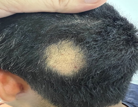
A non‑scarring patch measuring approximately 5 × 6 cm was seen in the occipito‑parietal area with sharp borders. No marked inflammation, infiltration, or atrophy.
Trichoscopy showed:
- reduced hair density;
- empty follicular openings;
- yellow dots;
- black dots;
- broken hairs;
- tapering (pencil‑point) hairs;
- short vellus hairs.
These findings are characteristic of alopecia areata and confirmed the diagnosis.
Fig. 1. Clinical appearance before treatment
Well‑defined non‑scarring patch in the occipito‑parietal area.
Fig. 2. Trichoscopy before treatment
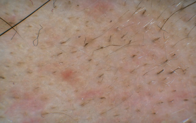
Yellow dots, black dots, broken and tapering hairs, and short vellus hairs.
Supportive laboratory tests
The diagnosis of alopecia areata was made clinically and trichoscopically. Additional tests were ordered to assess possible associated autoimmune/metabolic conditions:
- complete blood count;
- ferritin;
- TSH, free T4;
- anti‑TPO, anti‑TG;
- ANA;
- 25‑OH vitamin D;
- liver/kidney biochemistry when indicated.
These tests are not a specific biomarker for alopecia areata; they help rule in or out coexisting conditions (e.g., thyroid autoimmunity) and tailor treatment.
Treatment strategy
An individualized plan was built from clinical and trichoscopic activity, patch size, and duration.
Local therapy
- Intralesional triamcinolone acetonide into patches and margins every 4–6 weeks (first‑line for limited disease).
- Topical corticosteroid for 20 days initially.
- Calcineurin inhibitor after the steroid course to continue immunomodulation with less steroid burden.
- Minoxidil as adjunct therapy to support the anagen phase—not a primary immunologic treatment.
Systemic and adjunct care
- Methotrexate considered for multiple active patches, with clinical and lab monitoring.
- Zinc (50 mg) and vitamin D for documented deficiencies.
- PRP (platelet‑rich plasma) to support follicular recovery.
- Xelayt device as adjunct physiotherapy.
The patient was counseled that shampoo changes do not treat the underlying immune mechanism. Shampoo may support scalp hygiene but is not primary therapy.
Treatment course: six‑month follow‑up
Response was tracked clinically and with repeat trichoscopy—patch size, new hair growth, density, and trichoscopic activity.
After six months, clear improvement was documented:
- marked regrowth covering the patches;
- increased new hairs and improved density on trichoscopy;
- reduced activity of trichoscopic markers.
Fig. 3. Result after treatment
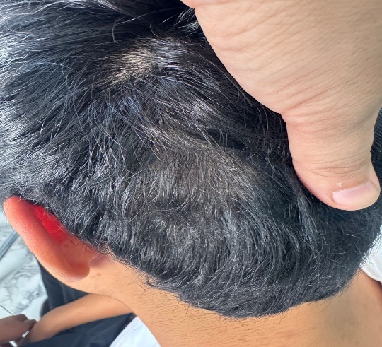
Hair regrowth in the alopecia patch after six months of combined treatment and follow‑up.
Conclusion
This case highlights timely diagnosis, trichoscopy, and individualized combined therapy in alopecia areata. Hair loss should not be dismissed as a “shampoo problem”—see a dermatologist or trichologist early.
Next steps at Radeski Skin Clinic
- Trichologist consultation.
- Computer trichoscopy.
- Personalized treatment plan.
- Ongoing clinical and trichoscopic follow‑up.
Related: Trichologist and trichoscopy and PRP for hair.
References
- Al-Dhubaibi MS, et al. Trichoscopy pattern in alopecia areata. Skin Research and Technology. 2023.
- Darwin E, et al. Alopecia Areata: Review and Treatment Options. Int J Trichology. 2018.
- Harries MJ, et al. BAD living guideline for alopecia areata 2025. Br J Dermatol. 2026.
Can hair loss be caused by shampoo?
Scalp hygiene matters, but alopecia areata is immune‑mediated. Shampoo changes do not treat the cause—do not delay a trichologist visit.
Why is trichoscopy needed?
It reveals characteristic signs (yellow/black dots, broken hairs) and objectively tracks treatment response.
Which blood tests are ordered?
As indicated—ferritin, TSH, vitamin D, autoimmune markers, etc.—to rule out associated conditions.
Can results appear in six months?
Outcomes vary. In this patient, combined therapy and follow‑up produced noticeable regrowth in six months; prognosis depends on activity and cause.