Surgitron Radiofrequency in Fergana and Kokand
Radiofrequency surgery — papillomas, molluscum and benign lesions with minimal scarring.
Surgitron RF provides gentle cutting and coagulation with minimal heat spread to surrounding tissue. At Radeski Skin Clinic papillomas, molluscum, keratomas and physician-approved benign lesions are removed; suited to delicate anatomical areas. Each lesion is assessed dermoscopically first.
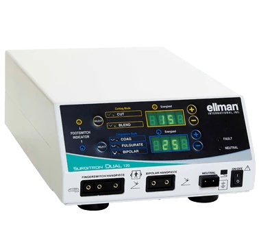
Surgitron is a radiofrequency surgery device made by the American company Ellman. It uses high-frequency radio wave energy to cut and coagulate tissue. Unlike a scalpel, it puts no mechanical pressure on the tissue, and unlike classic electrocautery, the electrode itself does not heat up: the heat is generated directly in the tissue cells.
In dermatology the device is used to remove benign skin growths: papillomas, soft fibromas (skin tags), molluscum contagiosum, keratomas and, when indicated, certain moles. Fine electrodes allow precise work even in delicate areas such as around the eyelids, the neck, the underarms and the groin.
At Radeski the key rule is that no growth is removed without being examined first. The dermatologist checks it with a dermatoscope and orders a histology test if there is any doubt. Tissue removed with the radio wave can be sent for histology when needed. If a malignant tumour is suspected, a different treatment approach is chosen and you may be referred to the appropriate specialist.
Manufacturer: Ellman (USA)
How does it work?
High-frequency current is delivered into the tissue through a fine electrode. The resistance of the cells turns this energy into heat: the water inside the cells evaporates instantly and the cells along the electrode's path separate. The cut is made with almost no pressure, and heating of the surrounding healthy tissue stays limited.
The device has several modes: cutting, cutting with coagulation, and coagulation. The doctor chooses the mode and electrode shape based on the type, size and location of the growth. Coagulation seals small blood vessels, so the procedure usually involves minimal bleeding, and the wound heals under a thin crust.
- Precise removal of the growth with a fine electrode
- Minimal bleeding thanks to simultaneous coagulation
- Limited heating of surrounding tissue
- The option of sending tissue for histology when needed
- Neat healing under a small crust
Indications
Papillomas and skin tags
Small hanging growths on the neck, underarms, under the breasts and elsewhere are often rubbed and injured by clothing. The radio wave removes them quickly and with minimal marks. Because papillomas can be linked to a virus, new ones may appear over time.
Molluscum contagiosum
Molluscum contagiosum is a viral skin infection that appears as small pearly bumps with a dimple in the centre. It is contagious and can spread across the skin. Removing the bumps helps limit the spread; the doctor chooses the treatment individually, especially for children.
Seborrhoeic keratoses
Keratoses are benign growths that appear with age, often brown with a rough surface. They can be removed if they bother you cosmetically or get caught on clothing. They can sometimes look like other lesions, so a dermatoscopic examination is essential.
Moles (naevi) when indicated
Benign moles that are repeatedly injured or bother you cosmetically are removed only after dermoscopy and with the dermatologist's approval. When needed, the removed tissue is sent for histology. Moles with signs of malignant change are not removed this way; a different approach is chosen for them.
Growths in delicate areas
Removing growths around the eyelids, on the neck, in the underarms and in the groin calls for particular precision. Fine electrodes and limited heat spread make careful work in these areas possible. In each case the doctor assesses at the examination whether the method is suitable.
Who is it for?
Radiofrequency surgery may suit patients who want growths that have been confirmed as benign removed quickly and with minimal marks. Before the procedure the doctor assesses:
- The nature of the growth based on dermoscopy
- The size, depth and location of the growth
- Whether you have a pacemaker or other implanted electronic devices
- Bleeding disorders and blood-thinning medications
- Allergy to local anaesthetics and how your skin heals
Contraindications
Whether the procedure is possible is decided individually. The main reasons to postpone removal or choose another method are:
- Suspicion of a malignant tumour: the growth is not removed this way until the diagnosis is clear
- An active infection or inflammation in the area, including a herpes flare-up
- A pacemaker or other implanted electronic device, which requires a separate assessment
- Bleeding disorders or anticoagulant use, where the risk is assessed in advance
- Fever, acute illness or uncontrolled chronic disease
- Conditions that impair healing, such as uncontrolled diabetes
- Pregnancy, when elective procedures are usually postponed and the decision is made individually
- Allergy to local anaesthetics, in which case another option is chosen
If you tend to form keloid scars, the risk of a mark is discussed separately. Papillomas and molluscum contagiosum are linked to viruses: removing a growth does not remove the virus itself, so new ones can appear over time.
How we use it at Radeski Clinic
Price list includes radiofrequency surgery positions — intradermal/epidermal lesions, complex zones and molluscum. Anaesthesia when needed is ordered by the physician.
Procedure steps
- Consultation and dermoscopy: The dermatologist looks at the growth and checks it with a dermatoscope, and asks about your medical history, medications and allergies. If there is any doubt, further tests or histology are arranged before removal. The method is chosen and the procedure explained at this stage.
- Anaesthesia: The area is cleaned with an antiseptic. Depending on the size and location of the growth, the doctor uses local anaesthesia, either an injection or a numbing cream. Very small growths sometimes do not need anaesthesia at all.
- Removing the growth: The doctor selects the right mode and electrode and either excises the growth or removes it layer by layer. Removing one small growth usually takes a few minutes. When needed, the removed tissue is sent for histology.
- Wound care: After the procedure the wound is treated with an antiseptic. Any minor bleeding is stopped with coagulation if needed. The doctor explains in detail how to look after the wound at home.
- Aftercare and follow-up: A small crust forms where the growth was and usually falls off on its own within one or two weeks. Do not pick the crust, rub the area or sunbathe until it has healed. If histology was done, you discuss the result with your doctor.
Results: what to expect
Once the crust falls off, a pink spot usually remains where the growth was, and over several weeks it gradually blends into the surrounding skin. How noticeable the mark will be depends on the depth of the growth, the area and your skin. You can expect:
- The growth gone, along with the discomfort it caused
- A small, neat mark in its place
- The pink spot gradually fading
- No more risk of the growth being caught on clothing
- If histology was done, a clear answer about what the growth was
Frequently asked questions
Is radiofrequency removal painful?
The procedure is done under local anaesthesia, so during removal you usually feel only slight pressure or warmth. The most uncomfortable moment is often the anaesthetic injection itself. Very small growths are sometimes removed without anaesthesia.
Will there be a scar?
Because the radio wave heats the surrounding tissue only slightly, the wound usually heals neatly. Whether a mark remains depends on the depth of the growth, the area, your skin and how well you follow the aftercare advice. It is important not to pick the crust and to protect the area from the sun.
Is it dangerous to remove a mole?
What is dangerous is removing a mole without examining it. That is why at Radeski every mole is first checked with a dermatoscope, and histology is ordered if there is any doubt. Moles with signs of malignant change are not removed with the radio wave; a different approach is chosen for them.
Can papillomas or molluscum come back?
A growth does not usually come back in the exact spot where it was removed, but because papillomas and molluscum are caused by viruses, new ones may appear elsewhere. If that happens, they can also be removed after a doctor's examination.
Can I get the area wet or go in the sun afterwards?
Your doctor will tell you exactly when you can get the area wet; rubbing or steaming the crust is usually not advised. Avoid sunbathing until the crust has fallen off and the skin has fully healed, and protect the pink spot with SPF for several weeks after that to lower the risk of a darker mark.
Conclusion
The Surgitron radiofrequency device is a way of removing benign skin growths precisely, with minimal bleeding and limited effect on the surrounding tissue. It is used to remove papillomas, molluscum, keratoses and, when indicated, certain moles, including in delicate areas.
At Radeski, removing any growth starts with a dermatologist's examination and dermoscopy. The doctor confirms that the growth is benign, orders histology if needed and chooses the most suitable method.