
Dermatopathology: Histology, Biopsy, and Tumor Diagnosis
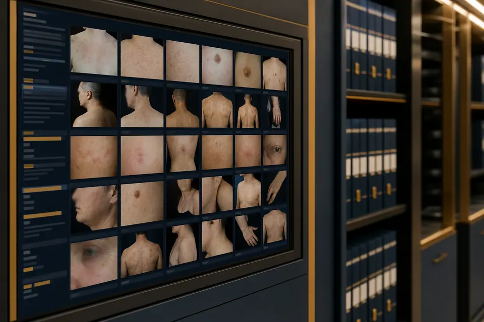
Dermatopathology: skin biopsy, histologic and immunohistochemical analysis, melanoma and tumor diagnosis — Radeski Skin Clinic capabilities.
Key takeaways
- Dermatopathology verifies diagnosis by microscopic study of skin samples
- Histology is the global standard when melanoma or tumors are suspected
- Radeski uses WHO, Breslow, Clark, and immunohistochemistry standards
- Both neoplastic and non-neoplastic skin disease is evaluated
- Pathologist and dermatologist jointly define the treatment plan
What is dermatopathology?
Dermatopathology is the laboratory specialty that examines skin biopsy samples under the microscope to verify the true nature of disease. Radeski Skin & Aesthetic Clinic has a dermatopathology department staffed by specialists trained at leading clinics in Russia and Europe.
The skin is the body's largest organ. Skin tumors such as melanoma are highly malignant and can metastasize early and extensively. Delayed or inadequate diagnosis can have serious consequences. That is why suspicious lesions require more than visual examination — histologic diagnosis is essential.
Why is histologic examination needed?
Many skin diseases share similar clinical features. The treating physician may be left with a picture that does not reveal the underlying process — and therefore cannot guide correct treatment.
Histologic diagnosis determines:
- whether a lesion is benign or malignant;
- the type of dermatosis;
- presence of infection (fungal, bacterial);
- changes in skin layers.
Today, skin histology is accepted worldwide standard practice. Modern surgical instruments allow samples to be obtained accurately and with minimal trauma.
What conditions are evaluated?
Neoplastic pathology
- nevi and other skin lesions (WHO and international classification);
- melanoma — Breslow, Clark, perivascular and perineural invasion, mitotic count, resection margins;
- basal cell and squamous cell carcinoma;
- Merkel cell carcinoma;
- adnexal skin tumors;
- soft tissue tumors;
- cutaneous B- and T-cell lymphomas (immunohistochemistry).
Non-neoplastic pathology
- various dermatoses;
- fungal and bacterial skin disease;
- photodermatitis;
- psychogenic dermatoses;
- hereditary, metabolic, and endocrine dermatoses;
- immune and connective tissue disorders;
- hair and nail disease.
These conditions are widespread and can significantly reduce quality of life. Accurate diagnosis is the first step to effective treatment.
Diagnostic capabilities at Radeski Skin & Aesthetic Clinic
- Punch biopsy analysis — special histochemical stains for fungal infection, dermal mucin, mast cells, elastic fiber disruption.
- Nevus and lesion diagnosis — reporting per WHO and international standards.
- Melanoma diagnosis — staging and prognosis per Royal College of Pathologists guidelines (Breslow, Clark, invasion, mitoses, margins).
- Sentinel lymph nodes — micrometastasis detection in melanoma by immunohistochemistry.
- Basal cell, squamous cell, and Merkel carcinoma — minimum dataset per Royal College of Pathologists.
- Adnexal skin tumors — hair, nail, and related structures.
- Soft tissue tumors — pathomorphologic evaluation.
- Cutaneous lymphomas — B- and T-cell markers, immunohistochemistry.
- Scientific collaboration — research integrated with clinical practice.
How does biopsy work?
Biopsy means taking a small tissue sample from a suspicious lesion. Steps:
- Examination by a dermatologist or dermato-oncologist.
- Dermoscopy if deeper assessment is needed.
- Sample taken under local anesthesia.
- Material sent to the laboratory.
- Microscopic analysis by a pathologist.
- Report and treatment plan.
Results are usually ready within several days; speed is especially important when malignancy is suspected.
Pathologist and dermatologist collaboration
At Radeski Skin Clinic, pathologist and dermatologist work together: clinical findings + laboratory report = accurate diagnosis. This approach is critical for melanoma, basal cell carcinoma, and complex dermatoses.
Related services: Dermatopathology, Skin biopsy, Dermoscopy, Dermato-oncology, Basal cell carcinoma article, Prices.
When to see a doctor
- A suspicious skin lesion has changed or is growing quickly
- Color, shape, or borders of a mole, spot, or sore have changed
- Biopsy was done but results and next steps are unclear
- Melanoma, basal cell carcinoma, or complex dermatosis is suspected
Frequently asked questions
Hashtags
#Dermatopathology#Histology#Skinbiopsy#Melanoma#Immunohistochemistry#RadeskiSkinClinic#Fergana
Important note: This article is for general information only and does not replace a medical consultation.
Related clinic services
More helpful articles
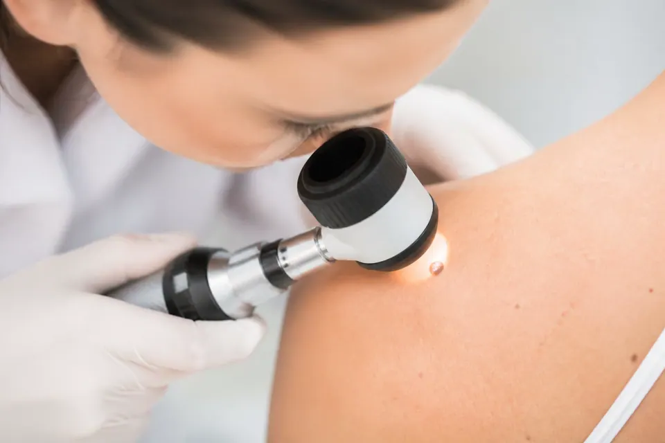
Moles and Dermoscopy: Which Mole Is Dangerous — the ABCDE Rule and Check-ups
When to check a mole: the ABCDE rule, risk groups, dermoscopy and mole mapping, removal and histology. Dermato-oncologists in Fergana and Kokand.
Read more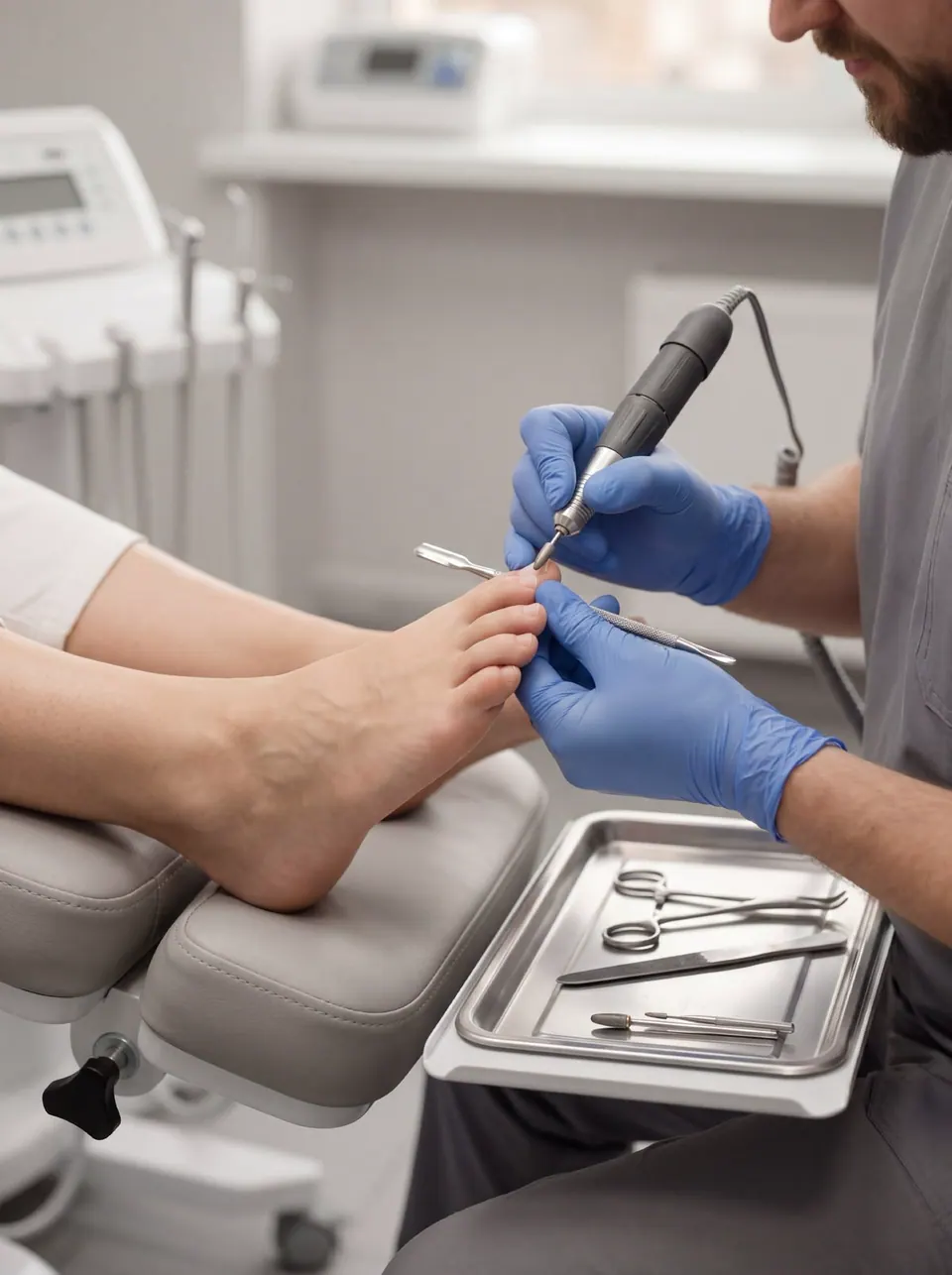
Nail Fungus (Onychomycosis): Signs, Causes and Treatment — Podiatrist Advice
Nail fungus (onychomycosis): yellow, thick, crumbling nails. Signs, lab confirmation, podiatric nail care and how long treatment takes. Podiatrists and dermatologists in Fergana and Kokand.
Read more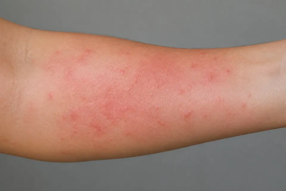
Skin Allergy and Hives: Symptoms, Causes and Treatment — When to Seek Urgent Help
Skin allergy and hives: itchy welts, redness and swelling. Causes, acute and chronic hives, when to call an ambulance and how it is treated. Dermatologists in Fergana and Kokand.
Read more